Welcome to the Department of Laboratory Animal Resources (DLAR) Imaging Core which provides imaging services for live animals, tissues and cells to support research performed at the University of Maryland, College Park campus. The core allows investigators to perform multiple imaging modalities which include the following equipment and Technology:
Fluorescent and chemiluminescent imaging utilizing the IVIS Spectrum In Vivo Imaging System
Rodent Ultrasonography utilizing the Vevo 3100 ultrasound unit
Rodent Computed Tomography with SPECT/PET capability utilizing the Albira Si CT-SPECT-PET unit
Radiography and fluoroscopic imaging using the OEC One Mobile C-arm
Multiphoton microscopy using the FVMPE-RS multiphoton laser scanning microscope
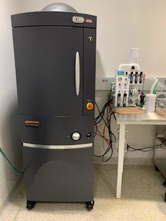
Irradiation service using the X-RAD 320 are also available
The A James Clark building also houses a state of the art vivarium and DLAR offices on the 6th floor. The vivarium contains rodent housing, large animal housing, procedure rooms, a dedicated surgery suite, cage wash support areas, and quarantine housing. DLAR oversees this facility and offers a variety of services including rodent housing and husbandry care, large animal housing and husbandry care, steam sterilization of materials, and ethylene oxide sterilization of materials. Surgery and procedure room space is available and anesthesia machines are available for use.
Compac5 Active Scavenging Mobile Multi-patient Anesthesia Unit for rodents
RC2 Passive Scavenging Portable Anesthesia Machines for rodents
Ethylene Oxide gas sterilization services for instruments and materials that cannot be steam sterilized
Steam sterilization services for surgical instruments and materials
Ionized hydrogen peroxide disinfection services using STERAMIST equipment
|
Hours |
Location |
|
Monday-Friday 8:30AM-4:30PM |
University of Maryland College Park 6th Floor, A. James Clark/Bldg. 429 8278 Paint Branch Drive College Park, MD 20742 |
 |
IVIS Spectrum In Vivo Imaging SystemThe IVIS Spectrum Imaging System enables high-sensitivity non-invasive in vivo fluorescent and bioluminescent imaging. This system features high resolution to 20 microns with a 3.9 cm field of view, high throughput imaging of up to 5 mice with a 23 cm field of view, and twenty eight high efficiency filters which span from 415-850nm for imaging across the blue to near infrared wavelength region. Spectral unmixing tools allow for the separation of signals from multiple fluorescent reporters within the same animal enabling the use of multiple reporters. An optical switch in the fluorescence illumination path allows illumination in either reflection-mode (epi-illumination/above) or transmission-mode (trans- illumination/below). Living Image Acquisition/Analysis Package software allows for advanced quantification and analysis of data. |
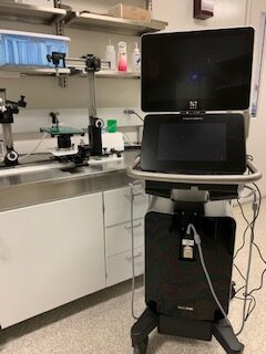 |
Vevo 3100 UltrasoundThe Vevo 3100 micro-ultrasound imaging system combines ultra high-frequency ultrasound imaging operating up to 70 MHz and imaging up to 300 frames per second. The system is equipped with complete animal setup and handling to achieve noninvasive in vivo imaging under accurate physiological conditions (temperature-controlled heated stage, gas anesthesia, with EKG, temperature, and respiratory rate monitoring). The system has an injection setup mounted on dedicated rail-system extensions to enable Y-axis stage adjustment that can be used to assist in image-guided injections. Three transducers are available: |
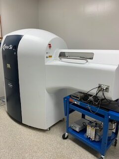 |
Albira Si PET/SPECT/CT imaging system The Albira Si is a trimodal imaging system that integrates Positron Emission Tomography (PET), Single-Photon Emission Computed Tomography (SPECT), and X-ray Computed Tomography (CT) within one fully x-ray shielded and stand-alone imaging platform. The fully automated animal handling system enables automatic co-registration of images. • Triple-ring Albira Si PET detector system with typical performance characteristics: FOV- transaxial 80mm, axial 148/286 mm (single position/moving table); spatial resolution up to 0.7 mm; sensitivity of 12%. |
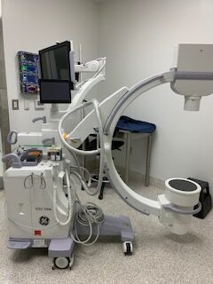 |
OEC One Mobile C-arm Fluoroscopic X-ray SystemThe OEC One Mobile C-arm system uses X-rays to provide fluoroscopic and digital spot/film images of the subject anatomy, surgical tools/devices, and/or contrast agents. The maximum field of view is 9 inches and maximal output is 110kVp/20mA |
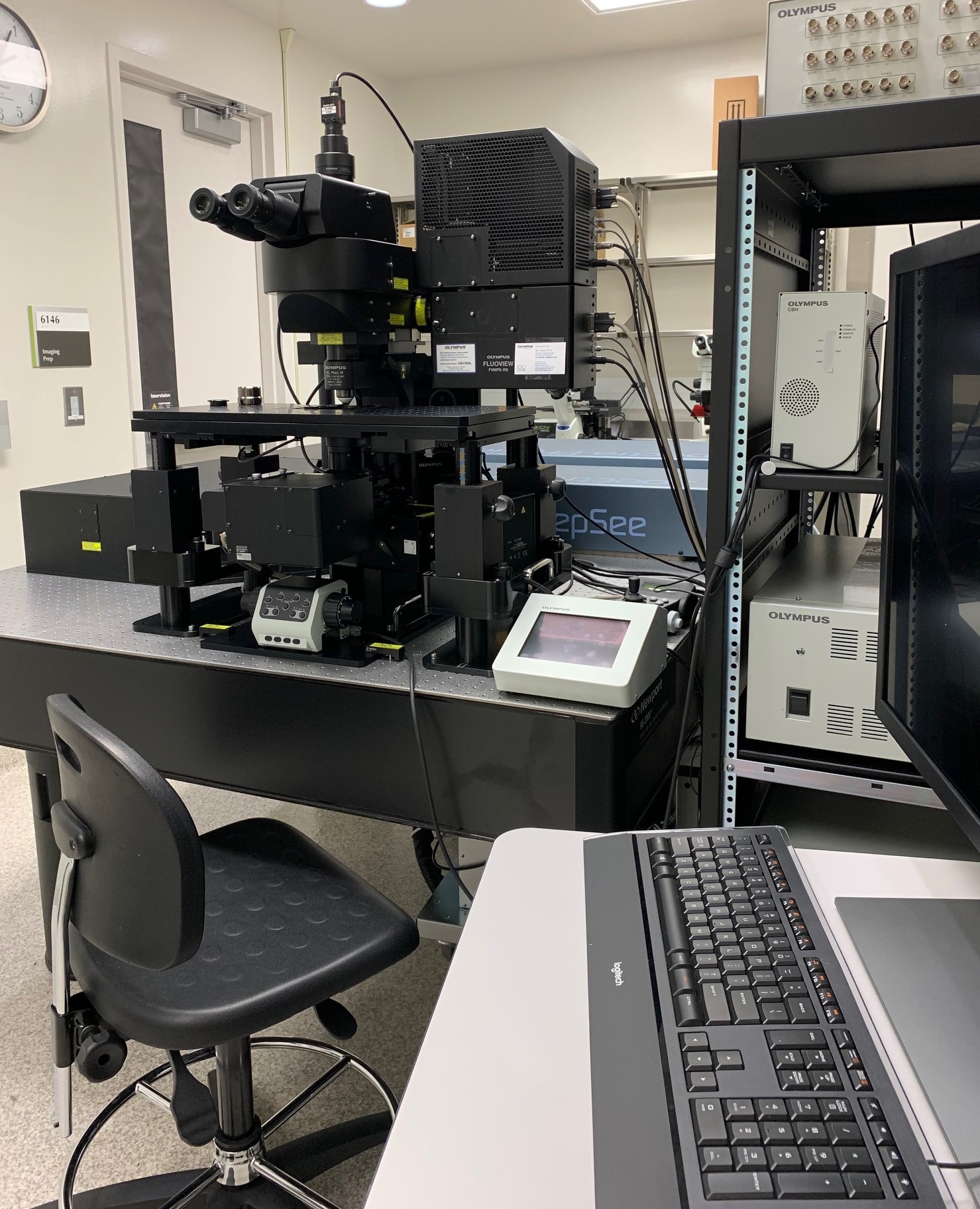 |
FVMPE-RS Multiphoton Laser Scanning MicroscopeThe FVMPE-RS multiphoton microscope employs advanced technology and optical design to enhance sensitivity and resolution during deep imaging of biological specimens.
The FVMPE-RS imaging platform supports a dual wavelength infrared pulsed laser or two independent tunable infrared lasers for multichannel, multiphoton excitation imaging. The dedicated high NA condenser detects transmitted fluorescence as well as transmitted second harmonic generation (SHG) signals. |
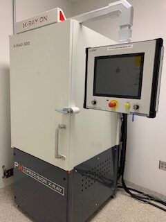 |
X-Rad 320 IrradiatorThe X-Rad 320 is a self-contained x-ray system designed to deliver a precise radiation dosage to biological samples including cell lines and rodents. The maximum output is 320kV with a maximum current of 30mA. The chamber contains an adjustable multi-position shelf with rotating platform to provide uniform radiation administration. • kV settings: 5kV-320kV in 0.1kV increments |
| Name | Role | Phone | Location | |
|---|---|---|---|---|
| DLAR Imaging Core |
DLARIC@umd.edu
|
6th floor A J Clark Hall
|
||
| Beth Bauer |
Imaging Core Director
|
301-405-6235
|
bbauer13@umd.edu
|
6106 A J Clark Hall
|
| Luis Lugo |
Attending Veterinarian
|
301-405-4920
|
llugo@umd.edu
|
6110 A J Clark Hall
|
| Betre Legesse |
Imaging Core Manager
|
301-405-5037
|
legesseb@umd.edu
|
6116 A J Clark Hall
|
| Jared Robinson |
Imaging Core Technical Coordinator
|
301-405-5498
|
jrobins4@umd.edu
|
6110 A J Clark Hall
|
| Service & price list |
| ► Miscellaneous (1) | |||
| Name | Description | Price | |
|---|---|---|---|
| DLAR - Sterilization Autoclave |
Internal
$28.35
each
|
||