Overview of Services

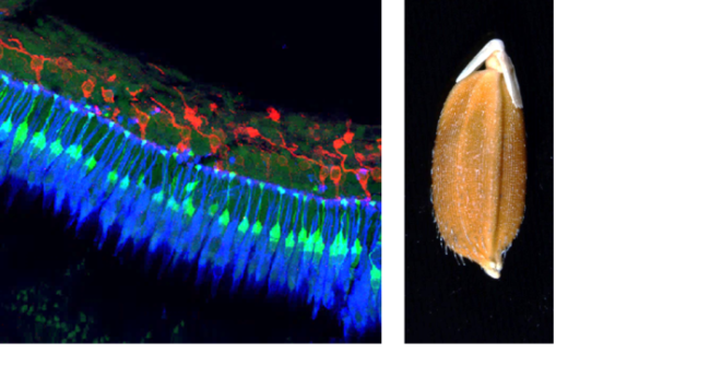
The Optical Imaging Core (OIC) provides detailed visualization of cell structure in biological processes through high-resolution imaging. Observation of development, pathogenesis, and genetic mutations at a cellular level allows investigators the opportunity to review how key components are organized, how they interact, and how they change in size and number. The fluorescent, confocal and multiphoton microscopes in the OIC are specifically designed for fluorescent imaging needed to gather this data. A variety of software programs are available for subsequent image analysis.
Flow cytometry is also available at the OIC. This provides cell characterization in large populations of dissociated cells ranging from bacteria to plant and mammalian cells. Flow cytometry is greatly enhanced by fluorescent labeling techniques, providing specific knowledge of gene, protein, and organelle changes within individual cells of a population. Once characterized, cells can be sorted for subsequent procedures (eg. incorporation of stem cells into animals or culturing of isolated bacteria) or analyses, such as proteomics and genomics. Cytometric analysis software is also available.
The OIC has a fluorescent stereoscope for imaging of plants and small animals that require a large depth of focus and wider field of view than can be provided in a standard compound light microscope.
The Optical Imaging Core is staffed by an experienced biologist and microscopist, Ann Norton.
Service Models
The Optical Imaging Core has two basic fee-for-service models.
The Self-Service Model is available to investigators who plan to acquire and analyze their own samples. Under this model, investigators will be billed for usage of instruments and training. It is the policy of the OIC that each user be trained by the OIC Director on instruments they plan to utilize. Once they are adequately trained, they will receive access to the OIC and will be allowed to schedule use of instruments independent of the OIC Director.
In the Full-Service Model, the OIC Director consults with investigators from experimental design through to publication of the study findings. The Principal Investigator or lab member will work closely with the OIC Director on protocols and timing of the samples. The OIC Director will acquire the images or cytometry data. Under this model, investigators will be billed for training, consulting, sample preparation, and usage of instruments.
The OIC Director Ann Norton can provide technical services, such as review and repair of microscopes outside the OIC, if time permits. Costs will be billed equivalent to consultation rates. Services provided by Ann:
Attribution
All papers, publications and press releases resulting from the use of the core facilities should include the following acknowledgement:
This project was supported by grants from the National Center for Research Resources (5P20RR016448-10) and the National Institute of General Medical Sciences (8 P20 GM103397-10) from the National Institutes of Health.
Leadership
Ann Norton, Director
asnorton@uidaho.edu
208-885-5191
Location
| Location | |
|
Life Science South 450 |
Links and Resources
| Name | Role | Phone | Location | |
|---|---|---|---|---|
| Ann Norton |
Director
|
208-885-5191
|
asnorton@uidaho.edu
|
LSS 450
|
| Optical Imaging Core Passes |
| ► Passes (3) | |||
| Name | Description | Price | |
|---|---|---|---|
| 3 Month Confocal Pass |
This pass allows trained users to use the Olympus Confocal Laser Scanning Microscope and the Nikon/Andor Spinning Disk Confocal Microscope. Usage time must continue to be logged, even though there will be no hourly billing. Limitations on this pass include: no more than 3 hours RESERVED/session, no more than 10 hours RESERVED/week for EACH PASS. Pass owners may use the instruments on the evening or weekends if no one has already reserved it. Compensation will not be offered for reasonable amounts of downtime. For longer time lapse imaging sessions on the spinning disk system, please contact Ann directly. |
Inquire | |
| 3 Month Flow Cytometry Pass |
This pass allows a trained user to do counting/analysis on the FACSAria Flow Cytometer. Sorting services on this instrument are only available on a Full Service basis (i.e. with the Director). Time on the instrument must continue to be logged even though charges will not be made on an hourly basis. Limits placed on this pass include: No more than 4 hours RESERVED/session; no more than 15 hours RESERVED/week; Compensation will not be offered for reasonable amounts of down time. If the instrument is unreserved in the evening or on weekends, a pass user can use it during that open time. |
Subscriber Price
$450.00
each
|
|
| 3 Month Non-Laser Microscopy and Analysis Pass |
This pass allows trained users to work on the Nikon Eclipse Fluorescent Microscope, the Leica MZ16 Fluorescent stereoscope, the Leica Histology microscope and any of the analysis computer stations. Again, to use any of these instruments you must be trained on each instrument by Ann, OIC Director. Policies associated with this pass include: no more than 4 hours RESERVED/session; no more than 15 hours RESERVED/week for each pass; hours of use must continue to be logged. Pass owners can use an instrument during the evening or weekend if no one has reserved it. |
Subscriber Price
$400.00
each
|
|
| Name | Price | |
|---|---|---|